4 Queen Anne St, London, England W1G 9ZF
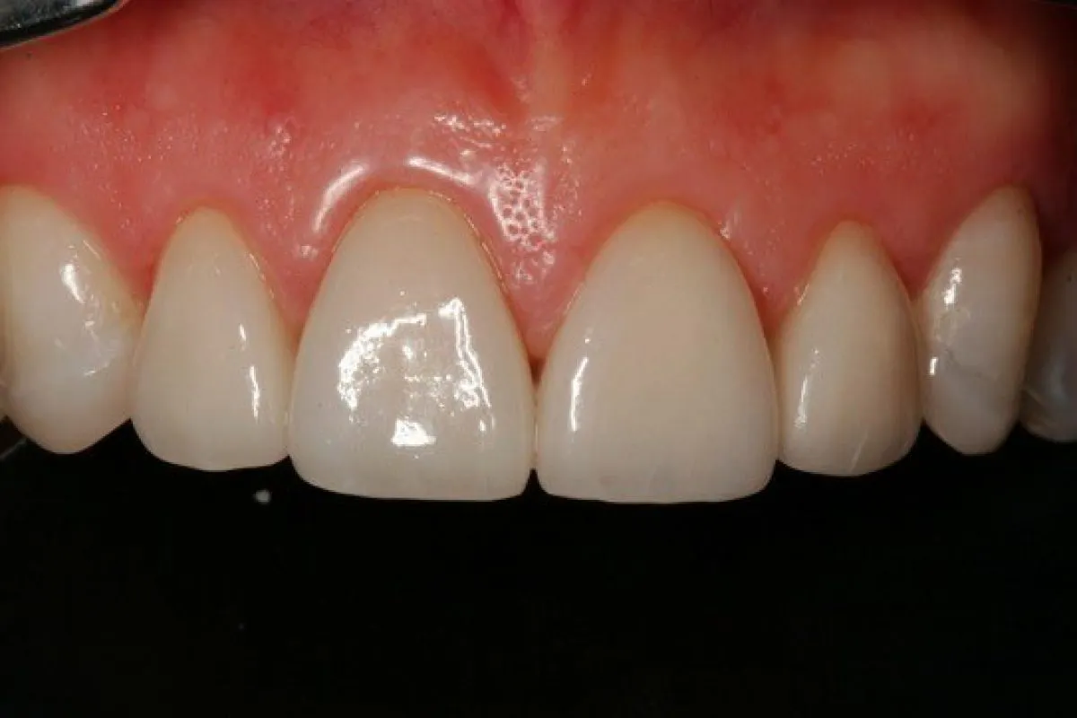
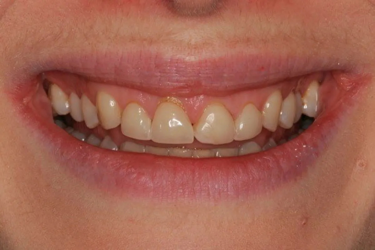
Simple gingivectomy can be performed if the tissue type is favourable and the excision does not encroach on the biologic width. In the case shown, the cemento-enamel junction could be felt probing buccally to confirm that the anatomical crown was the same length bilaterally. A peri-apical radiograph could also be used to confirm this. The excess tissue was removed by simple excision using a scalpel blade. A soft tissue laser or electrosurgery could also be used, but the wound is minimal and heals fully within days. There is a risk of the gum growing back, depending on the thickness of the tissue type and the position of the underlying alveolar bone. If this does occur, then crown lengthening utilising a full flap and bone removal will be needed for a stable result.
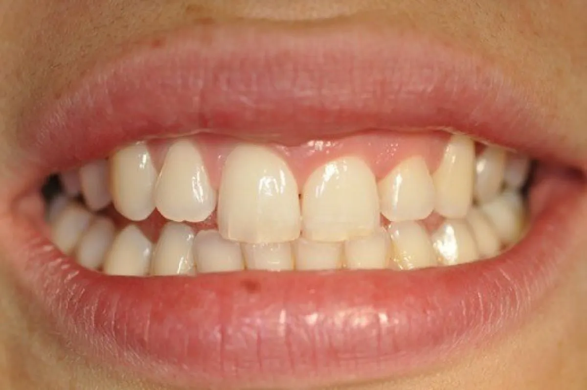
Before
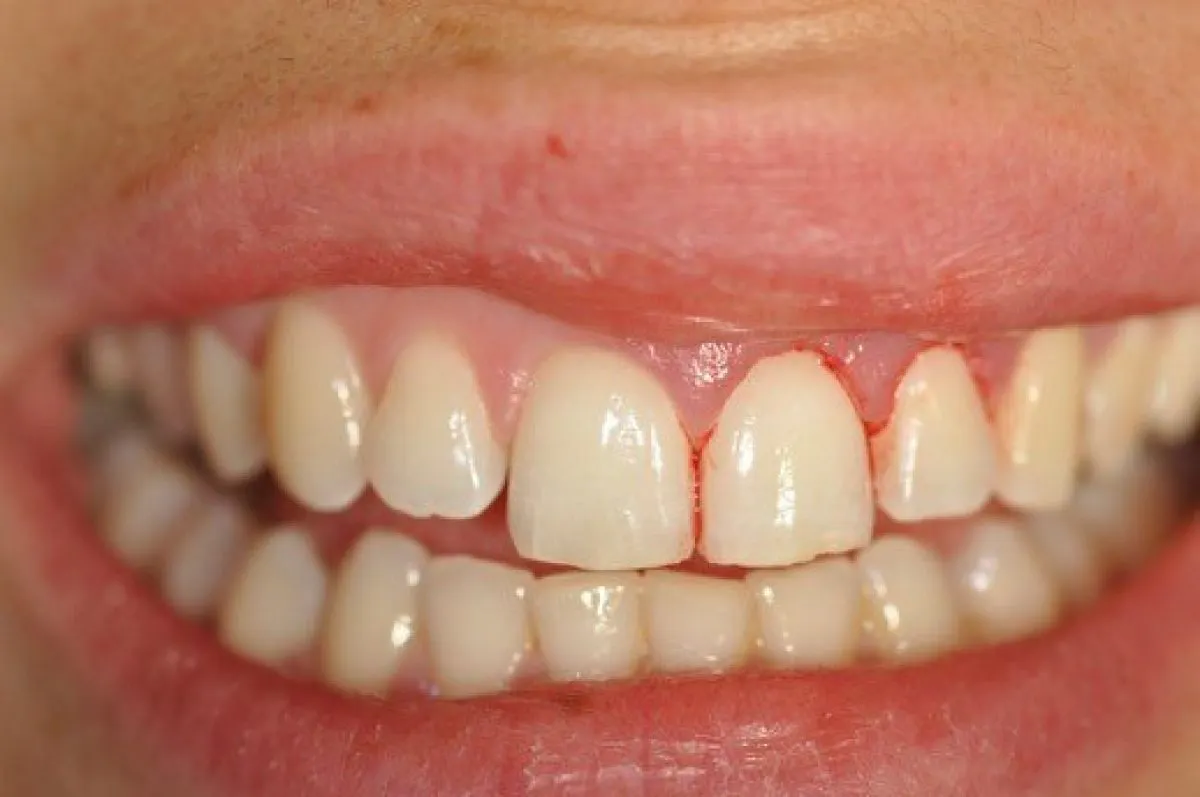
During
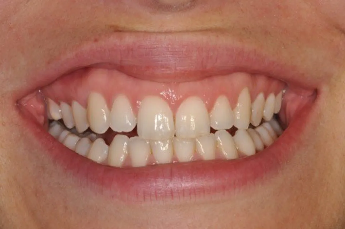
After
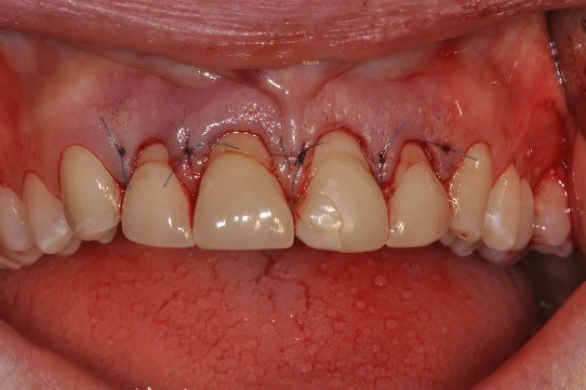
Surgical Crown Lengthening
A combination of tooth surface loss and an excess gingival display have left the patient with short teeth. The upper right central has some localized recession that puts its gingival margin to the same level as the upper canines. The decision was made to crown lengthen 12, 21 and 22 to match.
The initial incision removed the gingival tissue to the desired level. The flap is then joined up at the base of the papilla where there is sufficient width to eliminate the risk of losing any papilla height. Bone is then removed down to 3mm below the new gingival margin to establish a new ‘biologic width’ or to allow room for the reformation of a supracrestal attachment.
At 6 weeks the healing is reviewed and any small adjustments can be made by simple gingivectomy. Definitive restorations can be placed between 3 and 5 months depending on how invasive the surgery has been. (Restorations by Dr Robert Stone)
Before
During
After